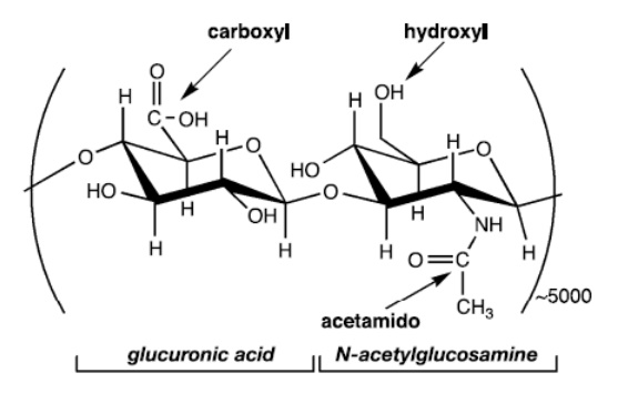
HA’s Complex Influence on Cell Migration

HA may influence cellular behavior not only through direct mechanisms (e.g., specific binding with cellular hyaladherins), but also by indirect means (through altering the physical properties of the ECM). HA’s effect on cell migration is a remarkable illustration of this complexity. For instance, the binding of cellular hyaladherins to HA is involved in migration for a variety of cell types. Yet, it is evident that HA may also indirectly aid cell migration by contributing to more open
and hydrated spaces in the ECM or by otherwise remodeling the environment through interactions with collagen and fibrin. Furthermore, as the main component of pericellular coats (matrices that are formed around migrating and proliferating cells), HA nonspecifically facilitates the detachment of cells from the ECM. Given
such complexity, distinguishing between the effects of HA’s biological activity and physicochemical properties is not straightforward.
HA Degradation
In mammals, the enzymatic degradation of HA results from the action of three types of enzymes: hyaluronidase (hyase), b-D-glucuronidase, and b-N-acetyl hexosaminidase. Throughout the body, these enzymes are found in various forms, intracellularly and in serum. In general, hyase cleaves high molecular weight HA into smaller oligosaccharides while b-D-glucuronidase and b-N-acetylhexosaminidase
further degrade the oligosaccharide fragments by removing nonreducing terminal sugars. Besides the degradation reaction, testicular hyase can catalyze the
reverse reaction, transglycosylation. Therefore, HA is not simply degraded by this enzyme into disaccharide fragments. On the contrary, incubation of HA in a testicular
hyase solution yields a mixture of fragment sizes, primarily made up of tetrasaccharides with smaller amounts of hexa-, octa-, and disaccharides. The cell
types primarily responsible for hyase synthesis are macrophages, fibroblasts, and endothelial cells; consequently, each of these cell types have been associated with
HA degradation in the body. In addition to enzymatic degradation, HA can also be degraded by reactive oxygen intermediates, a mechanism that has been implicated as a source of HA fragments at sites of inflammation. There are other nonenzymatic means of HA degradation(e.g., degradation induced by free radicals, ultrasound, pH, and temperature treatments).